Мышцы шеи
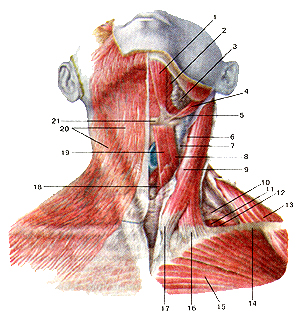
Рис.
110. Поверхностные мышцы шеи. Вид спереди. 1-переднее брюшко двубрюшной
мышцы; 2-челюстно-подья-зычная мышца; 3-подчелюстная слюнная железа;
4-шило-подъя-зычная мышца; 5-заднее брюшко двубрюшной мышцы;
6-внут-ренняя яремная вена; 7-обшая сонная артерия; 8-верхнее брюшко
лопаточно-подъязычной: мышцы; 9-грудино-ключич-но-сосцевидная мышца;
10-нижнее брюшко лопаточно-подъязычной мышца; 11-средняя лестничная
мышца; 12-задняя лестничная мышца; 13-трапециевидная мышца; 14-ключица;
15-6олыиая фудная мышца; 16-ключичная часть
фудино-клю-чично-сосцевидной мышцы; 17-фудинная часть
фудино-клю-чично-сосцевидной мышцы; 18-фудино-щитовидная мышца;
19-фудино-подъязычная мышца; 20-подкожная мышца шеи; 21-подъязычная
кость.
Fig. 110. Поверхностные мышцы шеи. Вид спереди.
1-m.digastricus (venter anterior); 2-m. mylohyoideus; 3-glandula
sub-mandibularis; 4-m.stylohyoideus; 5-m.digastricus (venter posterior)
; 6-vena jugularis interna; 7-arteria carotis communis; 8-m.
omohy-oideus (venter superior); 9-m. sternocleidomastoideus; 10-m.
omohy-oideus (venter inferior); 11-m. scalenus medius; 12-m. scalenus
posterior; I3-m. trapezius; 14-clavicula; 15-m. pectoralis major; 16-m.
sternocleidomastoideus (pars clavicularis); 17-m.
sternocleidomastoideus (pars sternalis); 18-m. sternothyroideus; 19-m.
sternohyoideus; 20-platisma; 21-os hyoideum.
Fig. 110. Superficial
muscles of neck. Anterior aspect. 1-anterior belly of digastric;
2-mylohyoid; 3-submandibular gland; 4-sty-lohyoid; 5-posterior belly of
digastric; 6-internal jugular vein; 7-common carotid artery; 8-superior
belly ofomohyoid; 9-sternocleidomastoid; 10-inferior belly of omohyoid
muscle; 11-scalenus medius; 12-scalenus posterior; 13-trapezius;
14-clavicle; 15-pectoralis major; 16-clavicular part
ofsternocleidomastoid; 17-sternaI part ofsternocleidomastoid;
18-ster-nothyroid muscle; 19-sternohyoid muscle; 20-platysma; 21-hyoid
bone.
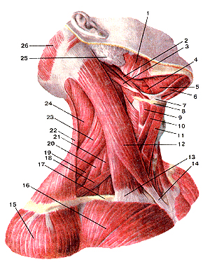
Рис. 111. Мышцы шеи. Вид справа.
I-жевательная
мышца; 2-шило-подъязычная мышца; 3-заднее брюшко двубрюшной мышцы;
4-подъязычно-язычная мышца; 5-челюстно-подьязычная мышца; 6-переднее
брюшко двубрюшной мышцы; 7-сухожильная петля, удерживающая сухожилие
двубрюшной мышцы возле подъязычной кости; 8-подьязычная кость;
9-щито-подъязычная мышца; 10-верхнее брюшко лопа-точно-подьязычной
мышцы; 11-фудино-иодъязычная мышца; 12-фудино-ключично-подъязычная
мышца; 13-ключичная головка фудино-ключично-сосцевидной мышцы;
14-грудинная головка фудино-ключично-сосцевидной мышцы;
15-дельтовид-ная мышца; 16-болыпая (рудная мышца; 17-ключица; 18-нижнее
брюшко лопаточно-подъязычной мышцы; 19-задняя лестничная мьммца;
20-средняя лестничная мышца; 21-передняя лестничная M,,;.iina;
22-трапециевидная мышца; 23-мышца, поднимающая лопатку; 24-ременная
мышца головы; 25-околоушная слюнная железа; 26-затылочное брюшко
надчерепной мышцы.
Fig. 111. Мышцы шеи. Вид справа.
1-m.masseter;
2-m.stylohyoideus; 3-m.digastricus (venter posterior); 4-m. hyoglossus;
5-m.mylohyoideus; 6-m.digastricus (venter anterior); 7-ansa tendinosus
dygastrici; 8-os hyoideum; 9-m.thyrohyoideus; 10-ven-ter superior m.
omohyoidei; ll-m. sternohyoideus; 12-m. stemocleidomastoideus;
13-capitulum claviculare m. sternocleidomas-toidei; 14-capitulum
pectorale m. stemocleidomastoidei; 15-m. del-toidens; 16-m. pectoralis
major; 17-clavicula; 18-m.omohyoideus (venter interior); 19-m.sca)enus
posterior; 20-m.scalenus medius; 21-m.scalenus anterior;
22-m.trapezius; 23-m.levator scapulae; 24-m.splenius capitis;
25-glandula parotidea; 26-m.occipitofrontalis (venter posterior).
Fig. 111. Masclesof neck. Right aspect.
1
-masseter; 2-styloihyoid; 3-posterior belly ol'digastric muscle;
4-hyo-glossus muscle; 5-mylohyoid; 6-anterior belly ol'digastric;
7-interme-diate tendon ol'digastric muscle is attached to hyoid bone by
a tascial loop; 8-hyoid bone; 9-stylohyoid muscle; 10-superiorbelly of
omohy oid muscle; I l-sternohyoid; 12- slernocleidomastoid;
13-clavicular head of sternocleidomastoid; 14-sternal head of
sternocleidomastoid; 15-deltoid; 16-pecloralis major muscle;
17-clavicle; 18-interior belly of omohyoid; 19-scalcniis posterior;
20-scalenus medius muscle; 22-scalenus anterior; 22-trapezoid;
23-levator scapulae; 24-spleniuscervi-cis; 25-parotid gland;
26-postcrior helly of occipitotrontalis.
7. Карыанный атлас
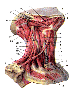 Рис. 112. Мышцы шеи. Подкожная мышца шеи и грудино-клю-
чично-сосцевидная
мышцы удалены. Вид справа. 1-челюстно-подъязычная мышца;
2-подъязычно-язычная мышца; 3-нереднее брюшко двубрюшной мышцы;
4-подъязычная кость; 5-щито-подъязычная мьшща; 6-НИжниЙ сжиматель
(констриктор) глотки; 7-всрхнее брюшко лопаточно-подъязычной мышцы;
8-грудино-подъязычная мышца; 9-грудино-щитовид-ная мышца; 10-щитовидная
железа; 11-пищевод; 12-трахея; 13-ключица (отрезана); 14-1-е ребро;
15-паружная межреберная мышца; 16-задняя лестничная мышца; 17-средняя
лестничная мышца; 18-передняя лестничная мышца; 19-нижнее брюшко
лопаточно-подъязычной мышцы; 20-длинная мышца шеи; 21-мышца,
поднимающая лопатку; 22-длинная мышца головы; 23-полуостистая мышца
головы; 24-длиннейшая мышца головы; 25-грудино-ключично-сосиевидная
мышца (отрезана); 26-заднее брюшко двубрюшной мышцы;
27-шило-подъязычная мышца; 28-жевательная мышца; 29-шило-язычная мышца.
Fig. 112. Мышцы шеи. Подкожная мышца шеи и грудино-клю-чично-сосцевидная мышцы удалены. Вид справа.
(-т.
mylohyoideus; 2-m. hyoglossus; 3-m.digastricus (venter anterior); 4-os
hyoideum; 5-m.thyrohyoideus; 6-m. constrictor pharyngis interior; 7-m.
omohyoideus (venter superior); 8-m. sternohyoideus; 9-m.
ster-nothyroideus; 10-glandulathyroidea; 11-esophagus; 12-trachea;
13-clav-icula (отрезана); 14-costa 1; 15-m. intercostalis externus;
16-m. scalenus posterior; 17-m.scalenusmedius; 18-m. scalenus anterior;
19-m. omohyoideus (venter inferior); 20-m.longus colli; 21-m. levator
scapulae; 22-m. longus capitis (отрезана); 24-m.semispinalis capitis;
24-m.longissimus capitis; 25-m. stemocleidomastoideus; 26-m.digastricus
(venter posterior); 27-m.stylohyoideus; 28-m.masseter;
29-m.styloglossus.
Fig. 112. Muscles of neck.
Platisma and
sternocleidomastoid muscle are cut away. Right aspect. 1-mylohyoid;
2-hyoglossus; 3-anterior belly of digastric muscle; 4-hyoid bone;
5-thyrohyoid muscle; 6-inferior constrictor (of phar ynx); 7-superior
belly of omohoid; 8-sternohyoid; 9-sternotliyroid; 10-thyroid gland; !
I-oesophagus; 12-trachea; 13-clavicle (cut away); 14-first rib;
15-external intercostal; 16-scalemis posterior; 17-scalenus medius;
18-scalenus anterior; 19-inferiorbellyofomohyoid; 20-longus colli:
21-levator scapula; 22-longUS capitis; 23-scrnispinalis capitis;
24-[ongissimus capitis; 25-sternocleidomastoid (cut away); 26-posterior
belly of digastric; 27-stylohyoid muscle; 28-masseter; 29-styloglossus.
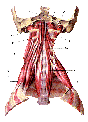
Рис.
113. Глубокие мышцы шеи. Вид спереди. 1-передняя прямая мышца головы;
2-боковая (латеральная) прямая мышца головы; 3-межпоперечные мышцы;
4-длинная мышца головы; 5-длиниая мышца шеи; 6-1-е ребро; 7-задняя
лестничная мышца; 8-средняя лестничная мышца; 8-передняя лестничная
мышца; 10-мьшща, поднимающая лопатку (отрезана); 11-11 шейный позвонок;
12-поперечный отросток атланта; 13-шиловидный отросток; 14-основная
(базилярная) часть затылочной кости.
Fig. 113. Глубокие мышцы шеи.
Вид спереди. 1-m. rectus capitis anterior; 2-m. rectus capitis
lateralis; 3-mm. inter-transversarii anteriores cervicis; 4-m. longus
capitis; 5-m. longus colli; 6-costa 1; 7-m. scalenus posterior; 8-m.
scalenus medius; 9-m. scalenus anterior; 10-m. levator scapulae
(отрезана); 11-axis; 12-processustransversusatlantis; 13-processus
styloideus; 14-parsbasilaris ossis occipitalis.
Fig. 113. Deep
muscles of neck. Anterior aspect, l-rectus capitis anterior; 2-rectus
capitis lateralis; 3-intertransversarii muscles; 4-longus capitis;
5-longus colli ; 6-first rib; 7-scalenus posterior; 8-scalenus medius;
9-scalenus anterior; 10-levator scapulae (cut away); ll-axis;
12-transverse process of atlas; 13-styloid process; 14-basal part
of"occipital bone. 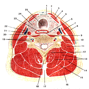
Рис.
114. Мышцы и фасции шеи на поперечном разрезе. 1-предтрахеальная
пластинка шейной фасции (средняя фасция шеи); 2-фудино-иодъязычная
мышца; 3-поверхностная пластинка шейной фаспии (поверхностная фасция
шеи); 4-грудино-щитовидная мышца; 5-подкожная мышца шеи;
6-предпозвоноч-ная пластинка шейной фасции (предпозвоночная фасция);
7-лопаточно-нодъязычная мышца; 8-грудиио-ключично-сосце-видная мышца;
9-длинная мышца шеи; 10-передняя лестничная мышца; 11-средняя и задняя
лестничные мышцы; 12-полуостис-тая мышца шеи; 13-полуостистая мышца
головы; 14-мышца, поднимающая лопатку; 15-ременные мышцы головы к шеи;
16-трапециевидная мышца; 17-выйная связка; 18-осгистый отросток шейного
позвонка; 19-позвоночные артерия и вена; 20-блуждаюший нерв; 21-общая
сонная артерия; 22-внутреняя яремная вена; 23-пищевод; 24-щитовидная
железа; 25-грахея.
Fig. 114. Мышцы и фасции шеи на поперечном
разрезе. I -lamina pretrachealis fasciae cervicalis;
2-m.sternohyoideus; 3-lam-ina superticialis fasciae cervicalis;
4-m.sternolhyroideus; 5-pla(isma; 6-lamina prevertebralis fasciae
cervicalis; 7-m. omohyoideus; 8-m. stern-ocleidomastoideus; 9-m. longus
colli; 10-m. scalenus anterior; 11-m. scalenus medius el m.posterior;
12-m. semispinalis cervicis; 13-m. semispinalis capitis; 14-m. levator
scapulae; 15-m. splenius capitis et m. splenius cervicis; 16-m.
trapezius; 17-lig. nuchae; 18-processus spinosus vertebrae cervicalis;
19-arteria vertebralis et vena vertebralis; 20-n. vagus; 21-arteria
carotis communis; 22-vena jugularis interna; 23-esophagus; 24-glandula
thyroidea; 25-trachea.
Fig. 114. Museles and faseias of neck
(horizontal section). 1-pretraeheal layer (fascia cerviealis);
2-sternohyoid; 3-superfacial layer (fascia cerviealis) investing layer;
4-sternotliyroid; 5-platysma; 6-prevertebral layer (fascia cerviealis);
7-omoliyoid; 8-sternocleidomas-toid; 9-longuscolli; 10-scalenus
anterior; I l-scalcni mcdiusand posterior; 12-semispinaliscervieis;
13-semispinaliscapitis; 14-levator scapulae; 15-splenius ccrvicis and
capitis; 16-lrapc/ius; 17-nuchal ligament; 18-spinous process of
cervical vertebrae; 19-vcrtebral artery and vein; 20-vagus nerve;
21-common carotid artery; 22-internal jugular vein; 23-oesophagus;
24-thyroid gland; 25-traehea.
ссылка - http://anatom.geiha.ru/data/12.htm
Источник: http://anatom.geiha.ru/data/12.htm | 